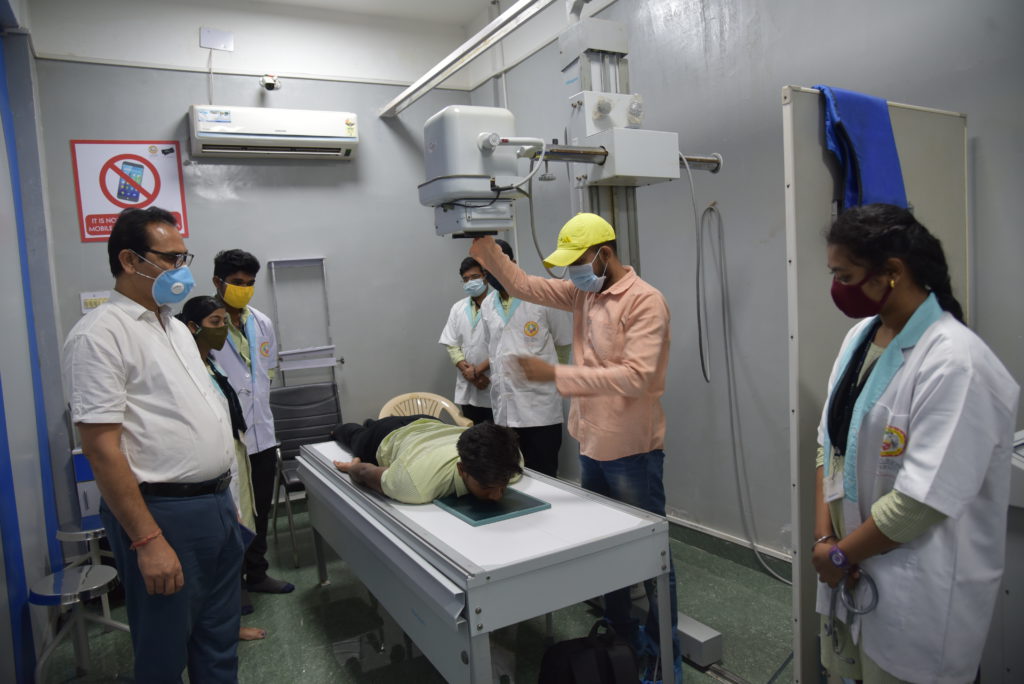
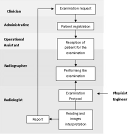
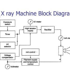
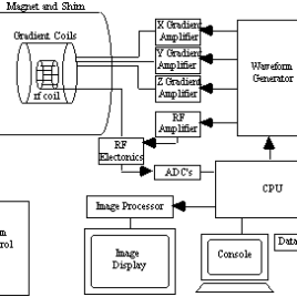
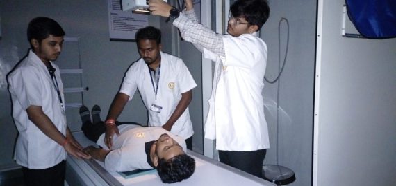
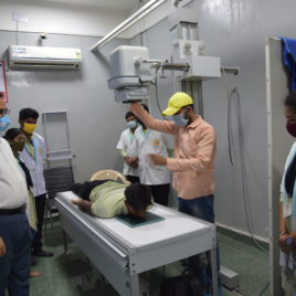
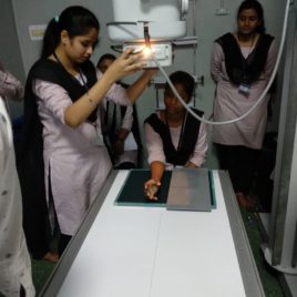
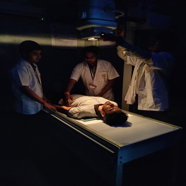
It’s a skill-oriented course to provide practice and knowledge to an individual to prepare the room and patient for performing diagnostic imaging examinations such as X-ray, CT scan and MRI under the guidance. Prepare the patients, unit & machine and keep patient records along with maintaining the standards of the equipment.
Radiology Technology
Course Attendees
Still no participant
Course Reviews
Still no reviews
Code(Credit) : SFS (0-2-2)
| Scheme | Skill for Success (SFS) |
| NSQF Level | 3 |
| Duration | 4 Months |
| Sector | Health |
| Occupations | Radiology Technician |
| Entry Qualification | XII |
| Minimum Age | 18 Years |
| Aligned to (QP) | HSS/Q0201 |
Course Objectives:
The objectives of this subject are:
- Covers the practical application of radiography in the paramedical profession.
- Includes principles of x-ray production, the operation and uses of x-ray machines, the care and development of films, and radiographic positioning of patients.
- Prerequisites: Admission to the X-Ray Technology program.
- Use an understanding of diagnostic quality images to determine options to correct deficiencies and to maximize diagnostic benefit and minimize radiation exposure based on repeated images.
- Apply knowledge of the health risks and effective safety procedures and professional judgment to minimize risks to personnel and patients during radiographic procedures.
After completing this program-
- The individual will be able to set up the X-ray, CT and MRI equipment’s to be used, ensure that safety precautions are taken to protect self, patient and staff from exposure to radiation
- Trainee will be expertise in preparing and positioning the patient correctly for different radiological procedures
- Able to prepare the room, apparatus and instruments for x-ray, CT scan, MRI
- Explain use of contrast material used for CT scan or MRI and how to administer them under supervision of the radiologist.
- Trainee will be able to manage a patient with contrast reactions and explain the use of protective materials
- Can assist in the department of Radio-diagnosis
- Can further pursue Diploma/ Degree courses in the same
Learning Record:
The trainee will submit a Practice/Project/Learning record after each class/session.
Assessment Process:
- The assessment of the theory/knowledge will be based on a written test/viva-voce or both, while the skill test shall be hands-on practical.
Course Syllabus/Contents:
Module I : Anatomy & Physiology
Skeletal System, .Respiratory system, Digestive System, Urinary system, Central nervous system, Endocrine system, Reproductive system.
Module II : Essential Physics
Sound & Heat, A.C. and D.C. power supply, Rectification and Transformers, Electromagnetic radiation spectrum and its properties, Production of X and gamma rays, Radioactivity, Interaction of radiation with matter, Thermoluminescent dosimeter (TLD), Radiation protection.
Module III : Principles of imaging
X-ray films, Intensifying screens, X-ray cassettes, Darkroom and equipment in Darkroom, Developer & fixer, Automatic Processing system, LASER Printer.
Module IV : Construction of Equipment
Stationery anode x-ray tube, rotating anode x-ray tube, Beam Restrictors, Grids, Fluoroscopy, Image Intensifier tube, Portable and mobile x-ray units, Dual-energy x-ray absorptiometry (DEXA).
Module V : Radiographic Technique– 1
- Upper limb, Lower Limb, Vertebral column, Skull Radiography, Chest X-Ray, Abdominal X-Ray. Views: AP, PA, LATERAL, OBLIQUE, RAO, LAO
Module VI : Radiographic Special Procedures
Contrast media used in the x-ray department (inclusive of those applicable to CT & MRI), Barium meal, Barium swallow, Barium enema, BMFT, IPV, HSG, RGU, MCUG, Myelography, Sialography, Angiography.
Module VII : Mammography & Ultrasound
Mammography Machine, Views, Ultrasound, Transducer, Probes, Properties of Ultrasound. Doppler Ultra Sound.
Module VII : Computed Tomography & Magnetic Resonance Imaging
History of CT & MRI, Generations of CT & Hounsfield Units, Concept of Voxel, Pixel, Basics of magnetism, Types of magnetism & Magnet, Larmour Frequency, Basic instrumentation of Coils, Advantages, Disadvantages, safety measures.
List of Projects/Products/Publications :
Reference Book :
- Basic Radiological Physics -K.Thayalan
- Radiological Procedure -Dr.Bhushan N Lakhar
- Manipal Manual of Anatomy -Madhyastha
- Clark's Positioning in Radiography-Hodder Arn
Session Plan:
Section 1
Anatomy & Physiology:
Skeletal System
Section 2
Module-1 Anatomy & Physiology
Topic : Respiratory System
Video :Section 3
Section 5
Section 6
Section 7
- Module-1 Anatomy & Physiology
Topic: Reproductive System
Video : - female-reproductive-system.mp4

- male-reproductive-system.mp4

Section 8
- Module-2 Essential Physics:Topic: IntroductionNotes : 2-Roentgen.pptVideo: X-RAY-INVENTION.mp4

Section 9
Module-2 Essential Physics:
Topic: Electromagnetic radiation spectrum and its properties, Production of X and gamma rays, Radioactivity, Interaction of radiation with matter
Notes : /Module-2-1.pptSection 10
Module -2 Essential Physics:
Topic: Thermoluminescent dosimeter (TLD), Radiation protection
Notes : TLD-BADGE.pptxSection 11
Module-3 Principles of Imaging

Topic : X-ray films, Intensifying screens, X-ray cassettes
Video : Adobe_Spark_Video.mp4Section 12
- Module-4 Construction Of Equipment
Topic : Stationery anode x-ray tube & Rotating anode x-ray tube
Video: - Adobe_Spark_Video-1.mp4

Section 13
- Module-5 Radiographic Technique -1
Topic : Upper Limb
Practice Video - HAND.mp4

- SHOULDER.mp4

- ELBOW.mp4

Section 14
- Module-5 Radiographic Technique -1
Topic: Lower Limb
Practice Video:
- KNEE.mp4

Section 15
Module-5 Radiographic Technique -1
Topic: Vertebral Column
Practice Video:Section 16
Module-5 Radiographic Technique -1
Topic : Chest X-Ray
Practice Video:Section 17
Module-5 Radiographic Technique -1
Topic: Abdominal X-Ray
Practice Video:Section 18
Module-5 Radiographic Technique -1
Topic: Pelvic Bone
Practice Video:Section 19
Module -6 Radiographic Special Procedures:
Topic: Barium Meal
Notes : Barium-meal/
Section 20
Module-6 Radiographic Special Procedures:
Topic : Barium Enema
Notes : Barium-enema/Section 21
Module-6 Radiographic Special Procedures:
Topic: Barium Meal Follow Through
Notes : Barium-meal-follow-through/Section 22
Module-6 Radiographic Special Procedures:
Topic: Barium swallow
Notes : Barium-swallowSection 23
Module -6 Radiographic Special Procedures:
Topic: IVP
Notes : Intravenous-urogramSection 24
Module-6 Radiographic Special Procedures:
Topic: MCUG
Notes: Micturating-cystourethrogramSection 25
Module - 7 Mammography and Ultrasound:
Topic: Mammography
Notes: Mammography
- Basic Radiological Physics -K.Thayalan
- Radiological Procedure -Dr.Bhushan N Lakhar
- Manipal Manual of Anatomy -Madhyastha
- Clark's Positioning in Radiography-Hodder Arnold
Industry Partnership :

Design Flow :
Latest News & Student Testimonials
CUTM3061 Testimonial | SKILL: Radiology Technician | CUTM
Media
Our Main Teachers

Sunil Kumar Jha is working as the Dean of School of Paramedics and Allied Health Sciences at Centurion University of Technology and Management since 2016. His major areas of expertise are medical diagnostics, clinical pathology, diagnostic bacteriology, histopathology and patient safety. He has completed MBA-HMGT, MLT, DAF, MHD and some of the courses in Alternative […]

